Read more
Your partner
in preventing infection.
Fast, safe, one-button
UV-C high-level disinfection for
ultrasound probes.
Our mission is to prevent healthcare-associated infections.
Healthcare-associated infections (HAIs) are a common and serious problem globally and locally. These infections are caused by cross-contamination of medical devices.
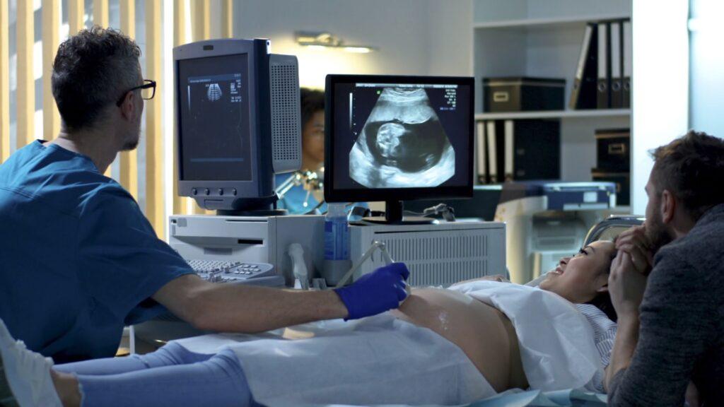
Women’s Health & IVF
13
%
of vaginal probes used with
sheaths test HPV positive after
disinfection with wipes.
Source: PLOS ONE
Cardiac Imaging
70
%
of HAIs can be prevented by implementing new infection
control interventions.
Source: WHO
ICU & Emergency
15
6
%
of patients in low- and mid-income countries will acquire at least one HAI during their hospital stay.
Source: WHO
Products
Chronos®
Fast, safe, simple ultrasound probe disinfection
technology
Yuvee®
Next-level
UV-C disinfection

Improve patient care and healthcare staff
safety while accelerating workflows.
Cutting-edge Innovation
Yuvee® technology is a leader in ultrafast, automated, high-level disinfection (HLD).
Trusted and recognised
We are globally recognised and known for excellence in scientific innovation.
Eco-conscious solutions
Our disinfection method doesn’t involve toxic chemicals — it’s safer for people and the planet.
Chemical-free sustainability

MEDIA
Get the latest news and information
Browse our articles, videos, and more to keep up with UV-C HLD news
Get the latest news
and information.
Browse articles, videos, and more to keep up with UV-C HLD news.

International Women’s Day: Working to Enhance Women’s Health
Lorem ipsum dolor sit amet, consectetur adipiscing elit. Suspendisse varius enim in eros.

Germitec: UVC high-level disinfection technology for ultrasound probes
Lorem ipsum dolor sit amet, consectetur adipiscing elit. Suspendisse varius enim in eros.
Read more
GE HealthCare Probe Compatible With Germitec UV-C HLD
Lorem ipsum dolor sit amet, consectetur adipiscing elit. Suspendisse varius enim in eros.
Read more

Fertility Exhibition – January 2024
Lorem ipsum dolor sit amet, consectetur adipiscing elit. Suspendisse varius enim in eros.
Read more

Unveiling Our New Website and Branding
Lorem ipsum dolor sit amet, consectetur adipiscing elit. Suspendisse varius enim in eros.
Read more

Sonosite Ultrasound Probes Declared Compatible With Chronos®
Lorem ipsum dolor sit amet, consectetur adipiscing elit. Suspendisse varius enim in eros.
Read more